Introduction
The Problem of Image Segmentation
Digital images surround us. From satellite imagery and autonomous vehicles to social media and medical diagnostics, the ability to extract meaningful information from visual data has become one of the defining challenges of the modern era. At the heart of this challenge lies a fundamental problem: image segmentation—the process of partitioning an image into distinct, meaningful regions. This book approaches image segmentation as a data science problem, drawing from the rich traditions of image processing while embracing the computational tools and methodologies that have transformed the field in recent years.
As Gonzalez and Woods articulate in their seminal treatment of digital image processing, segmentation algorithms broadly rely on two fundamental properties of image intensity values: discontinuity and similarity. Approaches based on discontinuity seek to partition an image by detecting abrupt changes—edges, boundaries, and contours that delineate where one region ends and another begins. Approaches based on similarity, by contrast, group pixels that share common attributes such as intensity, color, or texture. This book explores both paradigms, but does so through the lens of modern data science, treating segmentation not merely as an image processing task, but as a classification and optimization problem amenable to a diverse set of computational strategies.
The applications of image segmentation are vast and consequential. In remote sensing, segmentation enables the identification of land use patterns, deforestation, and urban expansion from satellite imagery. In autonomous driving, accurate segmentation of road scenes—distinguishing pedestrians from vehicles, lane markings from curbs—is a matter of safety and life. In manufacturing, segmentation supports quality control by identifying defects in materials and assemblies. And in medicine, segmentation forms the backbone of computer-aided diagnosis, enabling clinicians to identify tumors, measure organ volumes, and track disease progression with a precision that the human eye alone cannot achieve.
Our Focus: Urothelial Cell Nuclei Identification
Throughout this book, we anchor our exploration of segmentation methodologies in a specific, real-world application: the identification and delineation of nuclei from urothelial cell images. Urothelial cells line the urinary tract, and their microscopic examination plays a critical role in the detection and monitoring of bladder cancer and other urological conditions. The ability to accurately segment cell nuclei from surrounding cytoplasm and background is essential for downstream analysis—measuring nuclear size, shape, and chromatin texture—all of which carry diagnostic significance.
The ultimate aim of this segmentation task is not merely to identify where the nucleus resides within a cell, but to enable precise quantitative measurements. Chief among these is the nuclear-to-cytoplasmic ratio (N/C ratio)—the ratio of the area occupied by the nucleus to the area occupied by the cytoplasm. In healthy urothelial cells, the nucleus occupies a relatively small fraction of the total cell area, and the N/C ratio falls within a well-characterized normal range. However, in cells undergoing malignant transformation, the nucleus tends to enlarge disproportionately relative to the cytoplasm, resulting in an elevated N/C ratio. This biomarker is one of the key morphological indicators used by pathologists when evaluating cytology specimens for signs of dysplasia or carcinoma. An automated, reliable segmentation pipeline that accurately delineates both the nucleus and the cytoplasm therefore has direct clinical utility: it enables the computation of N/C ratios across large populations of cells, supporting early detection and risk stratification for bladder cancer and related conditions.
Consider the image below, which shows a urothelial cell alongside its corresponding segmentation target. The original cell image, captured under microscopy, presents a complex visual scene: the cell body appears in grayscale tones with varying intensity, and the nucleus—our region of interest—occupies a darker region within the cell. The target segmentation map, shown to the right, assigns distinct labels to the background (purple), the cell body or cytoplasm (teal), and the nucleus (yellow). With the nucleus and cytoplasm cleanly segmented, computing the N/C ratio becomes a straightforward matter of counting the pixels assigned to each region.
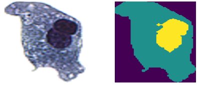
This seemingly straightforward task—identifying the nucleus within a cell—is in fact a microcosm of the broader segmentation problem. The boundaries between the nucleus and cytoplasm are not always crisp. Intensity gradients are subtle, noise corrupts the signal, and illumination varies from one specimen to the next. The challenge is to develop algorithms that can reliably and automatically perform this segmentation across thousands of cell images, with a level of consistency and accuracy that supports meaningful clinical analysis and accurate N/C ratio computation.
The Challenge of Noise and Ambiguity
The difficulty of the segmentation task becomes especially apparent when we examine cells where the visual distinction between nucleus and cytoplasm is less pronounced. Consider the second example shown below. Here, the cell appears as a roughly circular region of relatively uniform, dark intensity. The nucleus is embedded within this field, and the boundaries between nuclear and cytoplasmic regions are far from obvious to the naked eye. The corresponding segmentation target reveals the intended delineation, but arriving at this result algorithmically is a substantial challenge.
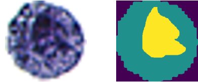
Images like this one illustrate why naïve approaches to segmentation often fail. Noise—whether arising from the imaging hardware, staining variability, or sample preparation—degrades the information content of each pixel. When the intensity difference between the nucleus and the cytoplasm is comparable in magnitude to the noise itself, simple thresholding rules break down. Edges become blurred, boundaries become ambiguous, and the segmentation problem demands more sophisticated solutions. It is precisely this progression—from simple to sophisticated—that forms the narrative arc of this book.
Images as Numbers: Exploring Pixel Data
To a computer, an image is simply an array of numbers. The urothelial cell image used throughout this book is stored as a NumPy array of shape (3 × 256 × 256)—three channels (red, green, blue), each holding 256 × 256 floating-point values between 0.0 and 1.0. This layout, called channels-first (or CHW format), is the default produced by PyTorch and many imaging libraries. Before we can index into it with the familiar image[row, col, channel] syntax, we transpose it to (256 × 256 × 3) — height, width, channels.
Use the explorer below to navigate the image with the row and column sliders, switch between color channels or grayscale, and inspect the raw float values behind each 5 × 5 patch. The Python code at the bottom updates live to show exactly how you would reproduce that view.
Methodologies at Our Disposal
The field of image segmentation offers a rich toolkit of methods, each with its own strengths and limitations. At the most fundamental level, intensity thresholding provides a direct approach: pixels above or below a chosen intensity value are assigned to different regions. While elegant in its simplicity, thresholding is highly sensitive to noise and assumes that regions of interest are separable in intensity space—an assumption that often fails in practice.
Beyond thresholding, we have access to a constellation of image processing techniques that serve as preprocessing steps, feature extractors, or standalone segmentation tools. Blurring and denoising operations, such as Gaussian filtering and median filtering, smooth out noise to reveal underlying structure. Edge detection algorithms—including Sobel, Canny, and Laplacian operators—identify regions of rapid intensity change that correspond to object boundaries. Morphological operations such as erosion, dilation, opening, and closing reshape binary regions to fill holes, remove small artifacts, and refine boundary contours. Region-based methods, including region growing and watershed algorithms, build up segmented areas by aggregating neighboring pixels that satisfy similarity criteria.
Each of these techniques addresses a different aspect of the segmentation problem, and in practice, effective segmentation pipelines often combine multiple methods in sequence. A typical workflow might begin with denoising, proceed through edge detection or thresholding, and conclude with morphological refinement. The art and science of image segmentation lies in selecting and tuning these operations for the problem at hand.
From Classical Methods to Modern Approaches
This book is organized around a progression of increasingly powerful approaches to solving the segmentation problem, using our urothelial cell dataset as a unifying thread.
Optimized Thresholding via Bayesian Methods. We begin by revisiting the classical method of intensity thresholding, but with a modern twist. Rather than manually selecting threshold values through trial and error—a tedious and subjective process—we frame threshold selection as an optimization problem. Using Bayesian Optimization, we systematically search the parameter space to find optimal or near-optimal thresholds that maximize segmentation quality with respect to a defined metric. This approach demonstrates how classical image processing techniques can be significantly enhanced by incorporating principled optimization strategies from the data science toolbox.
Classical Machine Learning. Our second approach departs from pixel-level intensity analysis and instead treats segmentation as a classification problem. Here, each pixel (or region) is classified into one of several categories—background, cytoplasm, or nucleus—based on a set of features. We explore two foundational machine learning algorithms for this task: k-means clustering, an unsupervised method that partitions pixels into groups based on similarity in feature space, and random forests, a supervised ensemble method that learns decision boundaries from labeled training data. These methods represent the bridge between traditional image processing and the data-driven paradigm that now dominates the field.
Convolutional Neural Networks. The most powerful approach to image segmentation in recent years has emerged from the field of deep learning, and specifically from convolutional neural networks (CNNs). CNNs have revolutionized computer vision by learning hierarchical feature representations directly from data, eliminating the need for hand-crafted features. For segmentation tasks, specialized architectures have been developed that can assign a class label to every pixel in an image—a task known as semantic segmentation. This book covers the fundamentals of neural networks and then focuses on the architectures that have proven most effective for our problem. We explore encoder-decoder architectures, in which an encoder (such as ResNet) progressively extracts high-level features through successive layers of convolution and pooling, while a decoder reconstructs the spatial resolution needed for pixel-wise classification. The U-Net architecture, originally developed for biomedical image segmentation, is given particular attention, as its skip connections between encoder and decoder layers enable the network to combine fine-grained spatial detail with high-level semantic information—a capability that is essential for accurately delineating structures like cell nuclei.
Structure of the Book
The book is designed as both a textbook and a practical guide, suitable for students and practitioners who wish to develop a deep understanding of image segmentation from first principles through to state-of-the-art methods. The progression of topics is deliberate, building from foundational concepts to advanced techniques.
We begin with an introduction to NumPy arrays—the fundamental data structure underlying all of our computational work. In the context of image processing and deep learning, these multidimensional arrays are often referred to as tensors, and developing a strong intuition for how to create, manipulate, and transform them is prerequisite to everything that follows. This foundational material covers array indexing, slicing, broadcasting, reshaping, and the basic arithmetic operations that are the building blocks of both image processing pipelines and neural network computations.
With this foundation in place, we proceed to image processing techniques using Python. Readers will learn to load, display, and manipulate images as arrays; convert between color spaces; apply spatial filters for blurring, sharpening, and edge detection; perform morphological operations; and implement basic segmentation algorithms. These skills are developed through hands-on examples and exercises, all implemented in Python using libraries such as NumPy, scikit-image, and OpenCV.
The first major project challenges the reader to perform segmentation of urothelial cell images using manual thresholding, followed by the optimization of thresholding parameters using Bayesian methods. This project bridges the gap between classical image processing and modern optimization, demonstrating that even simple algorithms can achieve surprisingly good results when their parameters are chosen wisely.
The second major project introduces classical machine learning approaches to segmentation, treating the problem as one of pixel classification. Through k-means clustering and random forests, readers learn to extract features, train classifiers, evaluate performance, and appreciate both the power and the limitations of these methods when applied to complex image data.
The final and most ambitious project tackles segmentation using convolutional neural networks. Before reaching this point, the book provides a thorough introduction to neural networks in general—covering perceptrons, activation functions, backpropagation, loss functions, and training procedures. We then specialize to the convolutional architectures that have become the gold standard for image analysis, culminating in the implementation and training of encoder-decoder and U-Net models on our cell image dataset.
The journey from a raw microscopy image to an accurate, pixel-wise segmentation map is a microcosm of the broader arc of modern data science: define a problem, understand the data, apply increasingly powerful tools, and measure the results. By the end of this book, readers will have not only a comprehensive understanding of image segmentation and its methodologies, but also the practical skills to implement, evaluate, and extend these techniques to new domains and new challenges. The urothelial cell serves as our guide, but the principles and tools developed here are broadly applicable wherever the extraction of meaningful structure from visual data is required.